The Body Palette: Your Inner Rembrandt
From my kitchen counter to Walt Whitman to Groucho Marx
The thin red jellies within you or within me, the bones and the marrow in the bones, / The exquisite realization of health; / O I say these are not the parts and poems of the body only, but of the soul.
— I Sing the Body Electric, by Walt Whitman
The Cut
Several years ago I sliced a finger joint right down to the bone, exactly where it bends. To be precise, that spot is called the distal interphalangeal joint — or the rather winsome “DIP” for short.
To briefly explain what happened, I was slicing some sweet potato on a newly acquired mandoline slicer. The tool came with a guard designed exactly to prevent this type of accident, but I stupidly decided that I was sufficiently nimble to not need that protection. Clearly a bad decision.
There was no pain. At least not at first. In fact, I didn’t even notice what had happened until I saw blood flowing down the mandoline and landing on the sliced potatoes. Red on orange.
Delayed pain reception is a documented phenomenon — not something I want to delve into in this essay. But it is relevant in that this pause of maybe a few minutes, in which I headed to the sink to begin running cold water over my finger and try to assess the damage, gave me the time to indulge in something I found quite curious.
The Beauty of Bone
I could clearly see the “live” bone, including the DIP joint. And its colour, to me, was a surprise. Perhaps like most people, I expected bone to be, well, “bone” coloured, like some designer wall paint. Or chalky white like skeletons. Hallowe’en-ish.
It wasn’t like anything I expected.
It was beautiful.
Opalescent. Iridescent. Shiny. Like mother-of-pearl. Pearlescent.
This was “live” bone, not some desiccated museum piece.
Despite being astonished by this unexpected beauty, I did need to stop the bleeding and attend to the minor medical emergency. Afterwards, I began to think about the wonder of the palette of the human body.
And, because my mind tends to spiral into odd corners of conjecture, I began to ponder whether a painter might consider using only colours found inside the human body.
Inner Beauty
In this mental exercise, I wanted to consider only “inner beauty” — in other words, I did not want to include exterior colours: no skin, no hair. For the same reason I have also set aside the eyes and the teeth, which, though technically visible only close-up, function more like exterior, on-display features than hidden interior ones. I wanted to focus only on healthy tissue, avoiding any colours associated solely with pathology, injury, or necrosis — just what is alive but ordinarily hidden from view.
And so, with this in mind, here is what turned out to be a surprisingly varied body palette.
Bone, Cartilage, and Tendon
Living cortical bone, freshly exposed, isn’t the chalky white of a dried anatomical specimen — that whiteness is a property of dead, degreased, bleached bone. Living bone has a blood supply running through it (via the Haversian canal system) and retains moisture and some organic content in its matrix, and the periosteum — the thin fibrous membrane covering bone — can itself have a glistening, slightly translucent quality. The gloss I noticed at the sink was this moist, vascularized surface catching light, the way any living tissue looks wet and lustrous compared to how we picture it dried out.
Living cortical bone runs pale ivory to warm grey-beige, with a faint sheen where the periosteum covers it. Cancellous (spongy) bone, exposed at a joint or fracture, is a deeper pink-tan, almost coral, saturated as it is with marrow and blood supply. Cartilage, often grouped with bone but structurally distinct, is a pale blue-white, almost translucent — think skim milk with a touch of grey, glossy like wet porcelain. Tendons are dense, fibrous, and pale ivory-white, with a slightly pearlescent sheen similar in tone to cartilage but less translucent.
Blood, and the Myth of Blue Veins
Blood itself, oxygenated or not, is never actually blue. Oxygenated (arterial) blood is bright, almost cherry red; deoxygenated (venous) blood is darker, more brownish-red or maroon — closer to an oxblood tone. What makes veins appear blue through skin is an optical effect rather than a pigment: skin and fatty tissue scatter and absorb red wavelengths of light more than blue, so blue-violet wavelengths preferentially scatter back to the eye from the light that reaches a vein and bounces out again.
A 1996 study using CCD imaging and Monte Carlo light-transport simulations confirmed that the perceived colour of a blood vessel depends on the scattering and absorption properties of the overlying skin, the oxygenation state of the blood, and the vessel’s depth and diameter — not on any blue pigment in the blood itself.1 It is the same broad category of phenomenon as why the sky is blue: light physics, not pigment.
Cut a vein open and drain it, and there is no blue to be found — just dark reddish-purple blood inside a translucent, greyish vessel wall.
There is, in short, essentially no true internal blue pigment anywhere in the body.
The one colour most people associate with the body’s interior in a sense of aristocracy is, on inspection, a colour that isn’t really there.
Muscle and Heart
Skeletal muscle is a deep, saturated red — the classic “raw meat” red — laced through and over with streaks of pale, silvery-white fascia. Heart muscle (the myocardium) is darker and more matte than most people expect, closer to a dried-blood or oxblood tone than to bright red. It is coated in a thin, glossy pericardial layer, with visible fat deposits along the vessels; that fat is a pale yellow-cream, a striking contrast against the dark muscle beneath it.
Nerve and Brain
Peripheral nerves are a distinctive pearly white to pale yellow-white, with a slightly glistening, cord-like appearance quite different from the surrounding tissue — part of why surgeons can identify them by sight alone.
The brain deserves special mention, because it corrects one of the most persistent misconceptions about the body’s colour palette. The living human brain is not grey. It appears as a soft pinkish-white, the result of constant blood flow through a dense capillary network; the familiar flat grey most people picture comes almost entirely from brains preserved in formaldehyde, which drains the blood and bleaches the tissue.7 In life, grey matter is genuinely pinkish-beige, and white matter is a brighter, cleaner off-white to yellowish-white, coloured by the fatty myelin sheathing its nerve fibres — the same fat-driven cream logic found elsewhere in the body’s fatty tissue.
The Digestive Organs
The liver is a deep reddish-brown, often described as maroon — one of the few organs to lend English a colour name in its own right. Its glossy outer capsule catches light with a wet sheen; the tone can shift toward purplish-brown when congested with blood, or toward tan-brown if fatty.
The stomach and intestines show two distinct faces. Their outer surface (the serosa) is smooth, glossy, pale pinkish-grey to tan, almost pearlescent. The inner lining (the mucosa), where visible, is far more vivid — deep pink to red, thrown into a rugose, folded texture that creates strong shadow and highlight, especially in the stomach. The small intestine’s interior runs a lighter, more coral-pink, with a velvety texture from its villi.
The pancreas is a pale, mottled yellowish-tan to pinkish-grey, lobulated and somewhat granular in texture, sitting stylistically between the pale tan of the kidney or spleen capsule and the cream of fat, since it is a gland embedded in and around fatty tissue. The kidneys are a deep reddish-brown, in the same family as the liver but usually a touch more purplish, with a smoother, tenser, glossy capsule.
Bile is genuinely one of the few sources of true green anywhere in the body — a dark olive-green to golden-green, sometimes shading toward brownish-green with concentration. The colour comes from bilirubin and its oxidation product biliverdin (literally “green bile”); bilirubin itself leans yellow-orange, and shifts toward biliverdin’s green as it oxidizes.2 Bile in the gallbladder runs darker and more concentrated than bile fresh from the liver, since the gallbladder actively reabsorbs water as it stores it.
The same green-yellow chemistry, running through the identical hemoglobin-breakdown pathway, is what colours a healing bruise as it moves from red-purple through blue-black to green and finally yellow-brown over one to two weeks — a visible, external timeline of an internal pigment process.
The Urinary System
The bladder wall, like stomach and intestinal serosa, is a smooth, glossy pale pinkish-grey on the outside. Its inner lining, when empty and relaxed, is thrown into folds with a pale pink to yellowish cast; when distended it stretches thin, paler still, sometimes showing a faint reddish tracery of small vessels.
And, I can personally attest to this, having had several cystoscopy procedures, where I was permitted to watch the “movie” from the camera’s perspective.
Urine’s colour comes from urochrome (urobilin), a downstream product of the same bilirubin pathway that colours bile — pale straw to deep amber depending on hydration and concentration. Urine and bile pigment are, in this sense, chemically related cousins, both tracing back to the breakdown of haemoglobin, presenting differently only because of where each sits in the degradation pathway.
The Reproductive Palette: Constant Change
The ovary is one of the most internally varied organs in the body’s whole palette, because its function is cyclical.
Its outer surface, the tunica albuginea (“white coat”), is a pale, dense, dull white-grey. After ovulation, the ruptured follicle transforms into the corpus luteum — literally “yellow body” — a saffron-yellow structure formed as the pigment lutein, a carotenoid related to the pigment in egg yolks and marigolds, accumulates in its cells.3 If no pregnancy follows, the corpus luteum degenerates into the corpus albicans (“white body”), a scarred, pale, fibrous remnant. Month after month, the ovary cycles between a white capsule, a saturated yellow secretory structure, and a fading white scar.
The uterus divides into three zones with three distinct colour logics. The serosa (outer covering) is smooth, glossy, pale pinkish-grey, in the same family as other abdominal organ coverings. The myometrium, the thick muscular wall, is a dense pinkish-tan to reddish-brown, muted compared to skeletal muscle, closer in spirit to heart muscle’s darker tone. The endometrium, the inner lining, is the single most dynamically coloured tissue in the whole survey: pale pink shortly after menstruation, thickening and growing markedly more vascular and deep red as it prepares for a possible pregnancy, before that same blood-rich layer sheds as the period.
The fallopian tubes are pale pink, delicate, and glossy, their small calibre giving an overall impression of soft coral rather than the deeper reds of larger organs; the fimbriae near the ovary run slightly deeper pink, owing to increased vascularity at that mobile end. The vaginal walls are pink to reddish-pink, thrown into folds like stomach or bladder mucosa, with a moist, glossy surface — one that shifts genuinely with hormonal state, thicker and more robustly pink under higher estrogen, paler and thinner in lower-estrogen states such as post-menopause.
In the male, the testes carry a pale bluish-white to greyish-white outer covering — the same tunica albuginea tissue type as the ovary’s capsule — with a softer, yellow-tan, coiled interior. The prostate is a pale pinkish-tan, firm glandular tissue, fairly nondescript, in the same family as the pancreas. Semen itself is a thick, whitish-gray fluid; a slight yellow tint is common and considered normal, varying with hydration and diet.4 It belongs to the same broad “milky, opalescent, warm-white” family as several other body fluids, since it is largely glandular secretion rather than a blood product.
The egg cell (oocyte) is unusual for being right at the edge of naked-eye visibility at all — roughly 100 to 120 micrometres across, making it the largest cell in the human body, at the very threshold of what the unaided eye can resolve.6 As for colour, it is generally described as transparent or colourless, without meaningful pigmentation, its appearance shaped more by light scattering and refraction than by any inherent hue — sometimes characterized as faintly yellowish or creamy under direct light. It is, chromatically, almost a null result: the one part of the reproductive palette defined by its near-absence of colour, in quiet contrast to the vivid cycling of the ovary that produces it.
Lungs, Glands, and Other Structures
Healthy lung tissue in a younger, non-smoking person is pale pink, almost salmon, mottled, spongy and matte rather than glossy. In practice, most adult lungs carry mottled grey-black patches from accumulated airborne carbon, creating a marbled pink-and-charcoal effect; the pleura, the outer lining, has a smooth, slightly glossy translucence over all of it.
The thyroid is a deep reddish-brown, highly vascular gland, closer in tone to liver or kidney than to the paler glands nearby. The adrenal glands are small but visually striking, with a genuine two-tone structure: a pale yellow outer cortex, rich in lipid content, surrounding a darker, greyish-tan inner medulla. The thymus, largest and most active in childhood, is a soft pinkish-grey; it shrinks and grows progressively more fatty and yellow with age, one of the few organs with such a marked lifetime colour shift.
The spleen is a deep purple-red to almost maroon-black, one of the darkest organs in the body, with a smooth, glossy capsule. Lymph nodes are small, pale pinkish-grey to tan, fairly nondescript and bean-shaped.
Fat deserves its own entry even though it is not an organ: pale yellow, sometimes almost cream, lobulated, matte rather than glossy, threading through and around nearly everything as a structural, connective colour.
Beneath the skin’s surface, the dermis itself carries a colour gradient rarely described in plain visual terms. The reticular layer — the deeper of the dermis’ two layers — is composed of dense, bundled type I collagen fibres, giving it a paler, denser, more fibrous ivory-white than the thinner, more vascular papillary layer above it, which is pinker and more blood-flushed.8 The reticular layer’s coarse, bundled collagen fibres are too large to produce the short-wavelength Rayleigh scattering that gives thinner, more disorganized tissue its blue-white translucence, so light reaching that depth is simply absorbed or scattered by whatever pigment — melanin, blood — happens to be present, rather than generating a scattering colour of its own.
Moving from the skin’s surface downward, the expected gradient runs from pink and vascular, to a denser pale ivory-white, to the cream-yellow of the fat layer beneath.
A Curious Aside: Earwax
Earwax is worth a brief detour, because its colour is one of the cleanest examples in the whole body of a single gene doing visible, direct work. People carrying one or two copies of a particular variant of the ABCC11 gene produce wax with higher fat content — wet, sticky, and brownish; those with two copies of the other variant produce wax with less fat — dry, light-coloured, and flaky. This single genetic variant was the first documented example of a DNA polymorphism determining a directly visible human trait5, a nice bit of trivia given how much messier most visible human traits are.
The Colours That Are Missing
Running this survey end to end, a pattern of absence is as interesting as the presence.
There is no true black under normal physiology. What reads as “black” in the body is almost always necrosis, pathology, or foreign material — dead or gangrenous tissue, very old oxidized blood, or the soot-like carbon deposits mottling a smoker’s lungs. True black is pathology or pollution, not baseline anatomy.
There is no pure, saturated orange. The nearest approaches are concentrated bile at certain stages of the bilirubin pathway, leaning orange-gold, and very concentrated urine running toward deep amber. Both are dilute or transient rather than a standing tissue pigment.
There is no true purple or violet. Venous blood leans purplish-maroon rather than true purple; the early stage of a bruise can look genuinely purple-black; the spleen, the closest standing candidate, still reads more maroon than violet.
There is no flat, opaque, printer-paper white. Bone, cartilage, tendon, and nerve are all pearled or ivoried, warm or faintly blue-white, but none is a true absolute white; the closest candidate, white matter in the brain, is only bright relative to the grey matter beside it.
And there is no vivid, saturated yellow of the egg-yolk or lemon variety — fat gives a pale cream-yellow, and concentrated bile or urine can push toward gold, but nothing internal reaches that fully saturated pitch, with the sole exception of the corpus luteum’s true saffron yellow.
The running theme: the body’s chromatic vocabulary is narrower and earthier than intuition suggests. Where popular imagination supplies bright orange organs, purple viscera, or black blood, it is drawing on illustration convention, decomposition, or disease — not living tissue.
An Ancestral Palette: Prehistoric Cave Art
This narrow, earth-toned range is not unique to the body. Prehistoric cave art’s palette is, almost by definition, constrained to nearly the same family of colours, and for a related reason: both are limited by what naturally occurring, unrefined materials can produce, without the technological capacity to synthesize or import brighter pigments.
Cave painters worked with red and reddish-brown from red ochre (hematite, an iron oxide), sometimes heated to shift the shade further toward red; yellow-brown from yellow ochre (goethite, a hydrated iron oxide); black from charcoal or manganese dioxide; and, where it appears at all, white from calcite, chalk, or kaolin clay.
Analysis of pigments from sites including Lascaux has confirmed that the reds are hematites and the yellows are goethites, with blacks consistently manganese-oxide based.9 Red ochre in particular sits in almost exactly the same chemical family — iron oxide — as blood’s colour and most of the body’s organ reds and browns, one from geology and one from biology.
What is conspicuously missing from the major Paleolithic sites is the same list as before: no blue, no green (with rare, debated exceptions), no true purple, no saturated orange beyond what ochre itself produces. Blue, in particular, had to wait for a real technological leap: Egyptian blue, generally credited as the first synthetic pigment, was in wide use by roughly 2500 BCE11, and lapis lazuli ultramarine came later still, remaining extraordinarily expensive for millennia.
Paleolithic painters were not choosing an earth palette aesthetically any more than the body “chooses” its own — both are working within a hard material constraint, one of available iron-oxide minerals and carbon, the other of available blood, bile, and connective-tissue chemistry, and both land in the same visual family as a result.
There is a further, more loosely evidenced layer worth flagging: a genuinely debated body of archaeological and ethnographic evidence suggests that some pigments, in some traditions, were mixed with blood, fat, or marrow as a binder, to help ochre adhere to rock surfaces.10
Where that happened, the connection between the body’s own palette and the cave painter’s palette stops being a mere resemblance and becomes, materially, the same substance.
This reframes the whole exercise: the “body palette” may not be an idle curiosity for a hypothetical modern painter so much as the default, ancestral human palette — the one our species used for tens of thousands of years before pigment chemistry expanded the available range. The bright, saturated palette most people now think of as “normal” for painting — vivid blues, clean greens, saturated purples — is the historical outlier, a comparatively recent expansion, not the baseline.
An Internal Palette
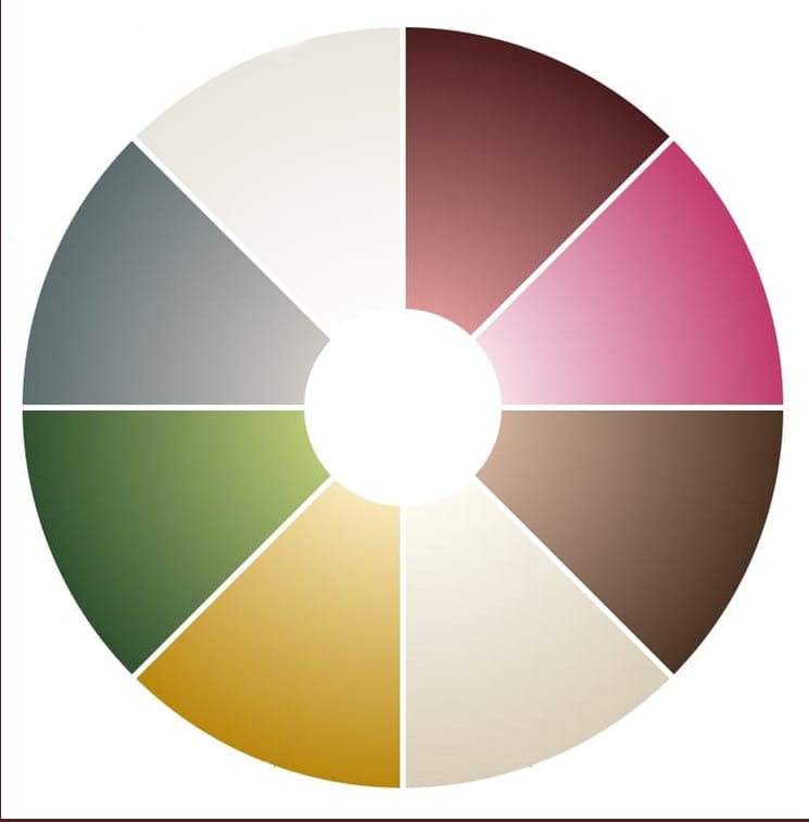
Gathered together, and excluding eye and tooth tones as set out at the start, here is the palette this survey turned up, grouped by family:
Reds
- Bright arterial crimson
- Deep venous plum
- Oxblood
- Cherry red
- Dusky brick red
Pinks & corals
- Pale rose pink
- Coral pink
- Salmon pink
- Vivid vascular pink
- Pinkish-beige
Browns & ochres
- Deep reddish-brown
- Maroon-brown
- Warm tan
- Greyish-tan
- Pinkish-tan
Creams & ivories
- Warm ivory
- Pearled off-white
- Blue-white
- Cream
- Pale straw-white
- Milky opalescent white
- Greyish-white
Yellows
- Pale cream-yellow
- Golden yellow
- Saffron yellow
- Amber
- Straw yellow
Greens
- Olive green
- Golden-green
- Dark bottle-green
Greys
- Soft grey-pink
- Ash grey
- Pale slate grey
- Grey-blue
- Clear / transparent
- Colourless, glass-clear
- Faint translucent white
A painter working strictly from this set would end up with something like a Rembrandt or Zorn-style palette naturally — dominated by reds, browns, and creams, with only a narrow, muted wedge of green, and no true saturated blue, black, purple, or orange at all.
Closing with Groucho Marx
It began at a kitchen sink, with cold water running over a cut I had no business getting, looking at a joint of my own finger and finding it, against every expectation, beautiful. Or, to close on a more irreverent note:
“Outside a dog, a book is man’s best friend; inside a dog, it’s too dark to read.”
— Groucho Marx
References
Cited works pertaining to colour and vision:
[1] Kienle, A., Lilge, L., Vitkin, I.A., Patterson, M.S., Wilson, B.C., Hibst, R., & Steiner, R. "Why do veins appear blue? A new look at an old question." Applied Optics, 35(7), 1151–1160 (1996).
[2] "Biochemistry, Biliverdin." StatPearls, National Library of Medicine (NCBI Bookshelf). https://www.ncbi.nlm.nih.gov/books/NBK549781/
[3] "Corpus luteum." Encyclopaedia Britannica. https://www.britannica.com/science/corpus-luteum
[4] "Semen: Fluid, Production, Storage & Composition." Cleveland Clinic. https://my.clevelandclinic.org/health/body/semen
[5] Yoshiura, K., Kinoshita, A., Ishida, T., et al. "A SNP in the ABCC11 gene is the determinant of human earwax type." Nature Genetics, 38, 324–330 (2006). https://www.nature.com/articles/ng1733
[6] "Egg comparison." National Institute of General Medical Sciences (NIGMS). https://nigms.nih.gov/image-gallery/1339
[7] "What are the Real Colors of a Human Brain? [The Living Truth]." academicpath.org. https://academicpath.org/real-colors-human-brain
[8] "Reticular layer of dermis" and "Papillary layer of dermis." Kenhub. https://www.kenhub.com/en/library/anatomy/reticular-layer-of-dermis ; https://www.kenhub.com/en/library/anatomy/papillary-layer-of-dermis
[9] "The raw materials." Lascaux cave, French Ministry of Culture. https://archeologie.culture.gouv.fr/lascaux/en/raw-materials
[10] "Stone Age Colour Palette: Paint Pigments Used in Caves." artslookup.com. https://artslookup.com/prehistoric/stone-age-colour-palette.html
[11] "Egyptian Blue Pigment Changes History." University of Memphis Media Room. https://www.memphis.edu/mediaroom/releases/2016/april16/egyptianblue.php